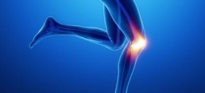
Osteoarthritis of the knee occurs when the cartilage in your knee joint breaks down, enabling the bones to rub together. The friction causes pain and the knee often becomes stiff and sometimes will swell. There are many treatments to slow its progress and ease your symptoms. Surgery is an option for more severe forms of osteoarthritis.
Cellstim Therapy Advanced Microcurrent delivers an automated multi-frequency waveform designed to accelerate healing through increased ATP production. A study of its effectiveness in treating osteoarthritis cartilage degeneration at the Canadian Memorial Chiropractic College indicated improved pain levels, knee joint function, and range of motion.
Case Studies: Osteoarthritis of the Knee
Case 1.
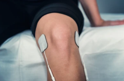
An 85 year old female with severe OA of the knee had been kept awake at night with pain. Prior treatments with anti-inflammatories and cortisone provided temporary, short term relief. CellStimTM treatment consisted of an IFC arrangement of pads on the medial/ lateral line and inf/ sup poles of patella. Treatments began at 3 times a week and then diminished. After 14 treatments in 10 weeks at 30 Hz/ 300 uA x 10 minutes and .3 Hz/ 40 uA x 10 minutes. The pain reduction indicated 95 % improvement.
Case 2.
A 70 year old male with moderate OA of the knee reports on occasion for treatment of knee pain. Previous treatment with IFC gave some improvement for short term relief. 20 – 30 minute CellStimTM treatments at .3 Hz and 40 uA biphasic gave lasting (2 weeks – 1 month) of significant relief (90-95% improvement). Pad placement was the same as case 1.
Case 3.
A 70 year old female with chronic OA of the 1st MTP was treated on 3 occasions with CellstimTM at .3 Hz at 30 uA biphasic once per week for 3 weeks. The joint was probed (using Qtip electrode probes) moving the probe location every 3 to 5 seconds. After 3 treatments the patient reported 90% improvement.
Chiropractic College … Canada
References
Effects of Electrical Stimulation (ES) on Articular Cartilage Regeneration with a Focus on Piezoelectric Biomaterials for Articular Cartilage Tissue Repair and Engineering January 2023
https://www.ncbi.nlm.nih.gov/pmc/articles/PMC9915254/
ES is a very effective stimulator to promote cartilage growth and repair. ES initiates cell differentiation by activating calcium transients to activate calmodulin and its downstream signals [95]. Studies have shown that ES reduces the level of type I collagen and increases the expression of chondrogenic markers, such as COL2α1, aggrecan, and Sox9 SOX9, thus promoting chondrogenic differentiation [96]. Ca2+ enters the cell primarily through voltage-operated calcium channels (VOCCs). Several studies have shown that the inhibition of VOCCs in stem cells leads to poor chondrogenesis, especially in the early stages [97]. This highlights an important role for VOCCs in cartilage development [98].
ES can not only activate ion channels but also some voltage-sensitive genes or receptors on cell membranes, thus initiating downstream signaling pathways and generating various biological reactions [84], mediating MSC condensation and chondrogenesis.
The CellStim waveform is effective for cartilage regeneration but will require several weeks of daily treatment twice a day. Long-lasting pain relief can be experienced within the first few treatments.
De Campos Ciccone C, Zuzzi DC, Neves LM, Mendonça JS, Joazeiro PP, Esquisatto MA. Effects of microcurrent stimulation on hyaline cartilage repair in immature male rats (Rattus norvegicus). BMC Complement Altern Med 2013 Jan 19; 13: 17. [DOI: 10.1186/1472-6882-13-17]
Abstract
Background: In this study, we investigate the effects of microcurrent stimulation on the repair process of xiphoid cartilage in 45-days-old rats.
Methods: Twenty male rats were divided into a control group and a treated group. A 3-mm defect was then created with a punch in anesthetized animals. In the treated group, animals were submitted to daily applications of a biphasic square pulse microgalvanic continuous electrical current during 5 min. In each application, it was used a frequency of 0.3 Hz and intensity of 20 μA. The animals were sacrificed at 7, 21 and 35 days after injury for structural analysis.
Results: Basophilia increased gradually in control animals during the experimental period. In treated animals, newly formed cartilage was observed on days 21 and 35. No statistically significant differences in birefringent collagen fibers were seen between groups at any of the time points. Treated animals presented a statistically larger number of chondroblasts. Calcification points were observed in treated animals on day 35. Ultrastructural analysis revealed differences in cell and matrix characteristics between the two groups. Chondrocyte-like cells were seen in control animals only after 35 days, whereas they were present in treated animals as early as by day 21. The number of cuprolinic blue-stained proteoglycans was statistically higher in treated animals on days 21 and 35.
Conclusion: We conclude that microcurrent stimulation accelerates the cartilage repair in non-articular site from prepuberal animals.
Treatment of Osteoarthritis of the Knee with pulsed Electrical Stimulation Thomas M. Zizic
Objective: The safety and effectiveness of pulsed electrical stimulation was evaluated for the treatment of osteoarthritis (OA) of the knee.
Methods: A multicenter, double blind, randomized, placebo controlled trial that enrolled 78 patients with OA of the knee incorporated 3 primary efficacy variables of patients’ pain, patients’ function, and physician global evaluation of patients’ condition, and 6 secondary variables that included duration of morning stiffness, range of motion, knee tenderness, joint swelling, joint circumference, and walking time. Measurements were recorded at baseline and during the 4 week treatment period. Results. Patients treated with the active devices showed significantly greater improvement than the placebo group for all primary efficacy variables in comparisons of mean change from baseline to the end of treatment (p <0.05). Improvement of > 50% from baseline was demonstrated in at least one primary efficacy variable in 50% of the active device group, in 2 variables in 32%, and in all 3 variables in 24%. In the placebo group improvement of > 50% occurred in 36% for one, 6% for 2, and 6% for 3 variables. Mean morning stiffness decreased 20 min in the active device group and increased 2 min in the placebo group (p <0.05). No statistically significant differences were observed for tenderness, swelling, or walking time.
Conclusion: The improvements in clinical measures for pain and function found in this study suggest that pulsed electrical stimulation is effective for treating OA of the knee.
Studies for longterm effects are warranted. (J Rheumatol 1995;22:1757-61)
In Vitro Growth of Bovine Articular Cartilage Chondrocytes in Various Capacitively Coupled Electrical Fields
Carl T. Brighton, Anthony S. Unger, and Jeffery L. Stambough Department of Orthopaedic Surgery, University of Pennsylvania School of Medicine, Philadelphia. Pennsylvania
Summary: Isolated articular cartilage chondrocytes from 1- to 3-week-old male Holstein calf knee joints were formed into pellets containing 4 x 10⁶ isolated cells and were grown in tissue culture medium (minimum essential medium/NCTC 135) containing either 1 or 10% newborn calf serum (NBCS) in plastic Petri dishes in 5% CO2 and air at 37 deg C in saturation humidity. On the 4th postisolation day either [3~S]sulfate or [3H]thymidine was added to the medium, and the pellets were exposed for 24 h to capacitively coupled electrical fields (10, 100, 250, and 1,000 V peak-to-peak, 60 kHz, sine wave signals). Current Intensity: 37 uA cm2 The pellets were then harvested, dialyzed, hydrolyzed, and assayed for DNA, protein, [35S]sulfate incorporation, and [3H]thymidine incorporation. Results indicated that at 250 V peak-to-peak there was a statistically significant increase in [35S]sulfate in 1% NBCS and a statistically significant increase in [3H]thymidine in 10% NBCS. At potentials above, or below 250 V no changes were noted. Thus, articular cartilage chondrocytes grown in pellet form can be stimulated to increase glycosaminoglycan synthesis or to increase cell proliferation by an appropriate capacitively coupled electrical field. The importance of the serum concentration in the medium in evaluation of biosynthesis in vitro is noted.

CellStim Therapy twice a day will provide long-lasting
pain relief and promote cartilage repair

Changing CellStim pad locations on each treatment increases effectiveness

